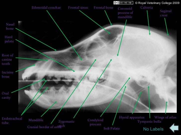
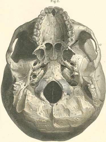
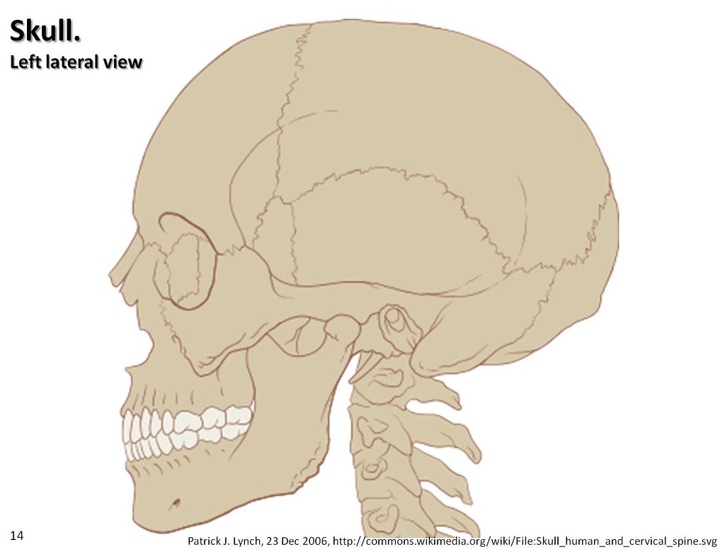
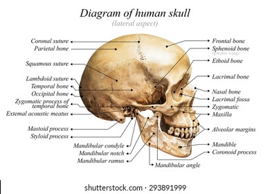
38 lateral view of skull with labels
Human penis - Wikipedia The human penis is an external male intromittent organ that additionally serves as the urinal duct.The main parts are the root (radix); the body (corpus); and the epithelium of the penis including the shaft skin and the foreskin (prepuce) covering the glans penis.The body of the penis is made up of three columns of tissue: two corpora cavernosa on the dorsal side and corpus … Clavicle (Collarbone) - Location, Anatomy, & Labeled Diagram 2. Acromial (Lateral) End. The broad, flat region of the clavicle lying towards the scapula is known as the acromial end or lateral end. It is both the widest and thinnest portion of the clavicle. This region bears a facet known as acromial facet that articulates with the scapula. The lateral end has two borders: anterior and posterior.
Nervous System – Medical Terminology for Healthcare Professions Labels read (top, left): pons, inferior olive, (top, right) cerebellum, deep cerebellar white matter (arbor vitae). In the top panel, a lateral view labels the location of the cerebellum and the deep cerebellar white matter. In the bottom panel, a photograph of a brain, with the cerebellum in pink is shown. [Return to Figure 8.7].

Lateral view of skull with labels
Skull Anatomy | The Neurosurgical Atlas The lateral surfaces of the labyrinth are covered by very thin, smooth plates called the lamina papyracea. 22 The posterior parts of the medial surfaces of the labyrinth contain thin, curved bones that form the superior nasal conchae and have an associated superior meatus. Anatomical Line Drawings - Medscape Spine - lateral view. go to drawing with labels go to drawing without labels. Lower Spine - anterior view. go to drawing with labels go to drawing without labels. Child Skeletal System - anterior ... (Solved) - Drag each label to the appropriate bone marking. Lines ... Drag each label to the appropriate ...
Lateral view of skull with labels. Part A Drag the labels onto the diagram to identify the bones and ... Part A Drag the labels onto the diagram to identify the bones and markings of the skull. Reset Help Foramen magnum Stylomastoid foramen Carotid canal External occipital protuberance IlII Occipital condyle Mastoid process Jugular foramen Exercise 9 Review Sheet Art-labeling Activity 4 Part A Drag the labels onto the diagram to identify the markings of the vertebral column. Wrist (lateral view) | Radiology Reference Article - Radiopaedia The academic rule of a true lateral wrist radiograph is defined by the pisoscaphocapitate relationship, where the palmar cortex of the pisiform should lie centrally between the anterior surface of the distal pole of the scaphoid and the capitate, ideally in the central third of this interval 1. Scapula - Parts, Anatomy, Location, Functions, & Labeled Diagram The scapula, alternatively known as the shoulder blade, is a thin, flat, roughly triangular-shaped bone placed on either side of the upper back. This bone, along with the clavicle and the manubrium of the sternum, composes the pectoral (shoulder) girdle, connecting the upper limb of the appendicular skeleton to the axial skeleton. Skull anatomy: Anterior and lateral views of the skull | Kenhub The bones of the skull that are visible from an anterior and a lateral view are the following: the sphenoid bone (with the greater and the lesser wings) the frontal bone (especially the orbital surface) the zygomatic bone the maxilla the mandible the nasal bones the ethmoid bones the parietal bone and the temporal bone
en.wikipedia.org › wiki › Human_penisHuman penis - Wikipedia The human penis is an external male intromittent organ that additionally serves as the urinal duct.The main parts are the root (radix); the body (corpus); and the epithelium of the penis including the shaft skin and the foreskin (prepuce) covering the glans penis. pressbooks.uwf.edu › medicalterminology › chapterNervous System – Medical Terminology for Healthcare Professions Anterior view labels indicate the right and left hemispheres and the longitudinal fissure between them. [Return to Figure 8.3]. Figure 8.4 image description: This figure shows the lateral view of the brain and the major lobes are labeled. From the front of the brain (left) labels read: frontal lobe, precentral gyrus, central sulcus, postcentral ... › en › e-AnatomyArm, forearm, and hand: MRI of anatomy - e-Anatomy - IMAIOS Sep 13, 2021 · By moving the mouse cursor over a particular area of the arm or forearm, this area is highlighted and the labels are displayed: anterior, lateral or posterior compartment. On the vertical left menu, a medical illustration of an upper limb skeleton based on a three dimensional (3D) model simplifies the access to the anatomical regions. Dog Skeletal Anatomy - Sheridan College 27/08/2022 · Acetabular Notch: Notch found on the ventral aspect of the acetabulum.; Acetabulum: A large articulation area with the head of the femur, and divided into Acetabular fossa, Lunatesurface.. Acetabular Fossa: A non-articular depression portion of the acetabulum used for the attachment of the ligament of the head of the femur.; Lunate Surface: Articular half …
A & P Unit 4 Flashcards | Quizlet inner surface of the skull. The subarachnoid space contains. cerebrospinal fluid (CSF) brain stem. Cerebellum. Cerebrum . Diencephalon. gray matter of spinal cord. gray matter of brain. Hypothalamus. Midbrain. occipital lobe. Olfactory tract of CN I. pineal gland. pituitary gland. postcentral gyrus. right cerebral hemisphere. substantia nigra. Thalamus. white mater of brain. … Cross-sectional anatomy of the brain - e-Anatomy - IMAIOS This module is a comprehensive and affordable learning tool for medical students and residents and especially for neuroradiologists and radiation oncologists. It provides access to an atlas and to images in axial planes, allowing the user to learn and review neuroanatomy interactively. Images are labeled, providing an invaluable teaching resource. Cerebral angiography - e-Anatomy - IMAIOS Brain - Angiography: Middle cerebral artery. Segments of the internal carotid artery (Bouthillier) - Angiography. Carotid bifurcation - Carotid sinus. Posterior cerebral artery - Anatomy (Angiography) Vertebral artery - Basilar artery:Cerebral angiography - Lateral view. Dural venous sinuses - Cerebral veins: Angiography - Lateral view. Forearm (lateral view) | Radiology Reference Article - Radiopaedia Forearm lateral view is one of two standard projections in the forearm series to assess the radius and ulna. Indications This view allows for the assessment of suspected dislocations or fractures and localizing foreign bodies within the forearm. Patient position patient is seated alongside the table
SNAP Tutorial and User's Manual SNAP represents segmentation by assigning labels to pixels (voxels) in the input image. For instance, when segmenting a brain MRI, some of the pixels in the image may be assigned the label 'grey matter', others will be assigned the label 'lateral vetricle', etc. It is up to the user to come up with the list of labels to use in a particular segmentation task. Each voxel in the input …

Dentistry lectures for MFDS/MJDF/NBDE/ORE: Diagrams Of Anatomy Of Skull With Radiographic Land Marks
quizlet.com › 538793548 › a-p-unit-4-flash-cardsA & P Unit 4 Flashcards | Quizlet inner surface of the skull. ... Identify the cranial nerves on this inferior-lateral view of a model brain showing blood supply. ... Place the following labels in the ...
Bone Is Composed of All of the Following Except Anatomyandphysiologytutor A Right Lateral View Of The Skull Complete With Labels Specifically Anatomy Bones Medical Anatomy Human Anatomy And Physiology Bone contains all of the following except _____. . If a bone is immersed in a weak acid such as vinegar for several days its inorganic components will dissolve.
› en › e-AnatomyAnatomy of the foot and ankle - MRI - e-Anatomy - IMAIOS Aug 26, 2022 · We used the Terminologia Anatomica to create the anatomical labels. This terminology is the international standard on human anatomy (it supersedes the previous standard, Nomina Anatomica since 1998). It was developed by the Federative Committee on Anatomical Terminology (FCAT) and the International Federation of Associations of Anatomists (IFAA).
Anatomy of the foot and ankle - MRI - e-Anatomy - IMAIOS 26/08/2022 · We used the Terminologia Anatomica to create the anatomical labels. This terminology is the international standard on human anatomy (it supersedes the previous standard, Nomina Anatomica since 1998). It was developed by the Federative Committee on Anatomical Terminology (FCAT) and the International Federation of Associations of Anatomists (IFAA). …
(Get Answer) - PART A: Assessments Identify the numbered bones and ... PART A: Assessments Identify the numbered bones and features of the skulls indicated in figures 14.30, 14.11. 14.12. and 1413 FIGURE 14.10 Identify the bones and features andicated on this anterior view of the skull using the terms provided if the line lacks the word bone, label the particular feature) FIGURE 14.11 Identify the bones and features indicated on this lateral view of the skull ...
Lights Holidays - See The Northern Lights In Lapland The Aurora Zone is THE original Northern Lights holiday company. We have built up an extensive range of trusted and knowledgeable guides, photographers and experts and we are quite simply your best chance to see the Northern Lights
Learn skull anatomy with skull bones quizzes and diagrams For this, take a look at our video below on the anterior and lateral views of the skull. Blank Skull Diagram Once you've done that, it's time to learn anatomy with our skull labeling quiz. We've created a blank skull diagram free for you to download as A PDF below. You can also download the labeled version and use this to make some notes.
Anatomy of the spine and back - e-Anatomy - IMAIOS Anatomical diagrams of the spine and back. This human anatomy module is composed of diagrams, illustrations and 3D views of the back, cervical, thoracic and lumbar spinal areas as well as the various vertebrae. It contains the osteology, arthrology and myology of the spine and back. It is particularly interesting for physiotherapists ...
vettech.sheridancollege.caDog Skeletal Anatomy - Sheridan College Aug 27, 2022 · The scapula is a flat triangular bone at the top of the shoulder; more commonly known as the shoulder blade. It consists of 2 surfaces (medial and lateral), 3 borders (cranial, caudal and dorsal) and 3 angles (craniodorsal, caudodorsal and ventral angle). The pig and horse do not have an acromion.
Label the landmarks of the skull in the figure below Skull is pictured from many angles. Skull Labeling - Answer Key. 1. Coronal Suture 2. Frontal 3. Parietal 4. Nasal 5. Squamosal Suture 6. Ethmoid 7. Lacrimal 8. Sphenoid 9. Lamdoidal Suture 10. Occipital 11. Temporal 12.. world war 2 worksheets pdf cute stories of how couples met english bedding mercedes comand knob not working
Posterior and lateral views of the skull: Anatomy | Kenhub Posterior and lateral views of the skull (overview) Sutures of the skull The lambdoid suture which can be described a non-movable fibrous joint which lies between the parietal and occipital bones. Its name derives from the Greek letter lambda which it is similarly shaped to.

Lateral View of the Sphenoid Bone | Neuroanatomy | The Neurosurgical Atlas, by Aaron Cohen-Gadol ...
The Brain and Nervous System | Noba The CNS is the portion of the nervous system that is encased in bone (the brain is protected by the skull and the spinal cord is protected by the spinal column). It is referred to as “central” because it is the brain and spinal cord that are primarily responsible for processing sensory information—touching a hot stove or seeing a rainbow, for example—and sending signals to …
Paranasal sinuses and facial bones (lateral view) - Radiopaedia The lateral paranasal sinuses and facial bones view is a nonangled lateral radiograph showcasing the facial bones (i.e. mandible, maxilla, zygoma, nasal, and lacrimal bone) and paranasal sinuses. Indications This view is useful in assessing any inflammatory processes or fractures to the facial bones, orbits, and paranasal sinuses.
Skull (lateral view) | Radiology Reference Article | Radiopaedia.org The skull lateral view is a non-angled lateral radiograph of the skull . This view provides an overview of the entire skull rather than attempting to highlight any one region. Indications This projection is used to evaluate for skull fractures, in addition to neoplastic changes and Paget disease.


Post a Comment for "38 lateral view of skull with labels"