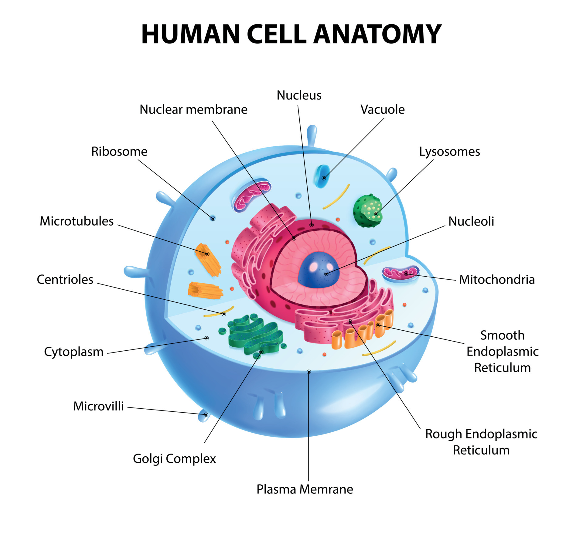
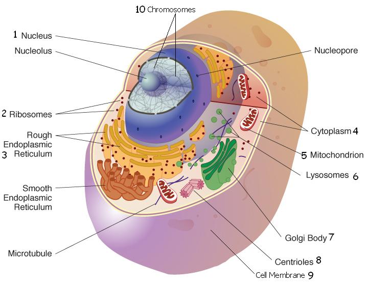
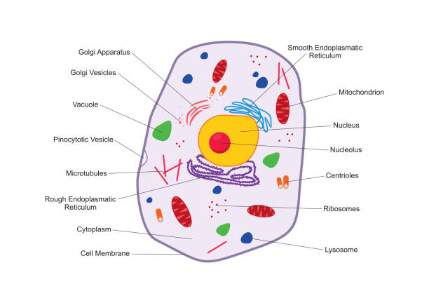
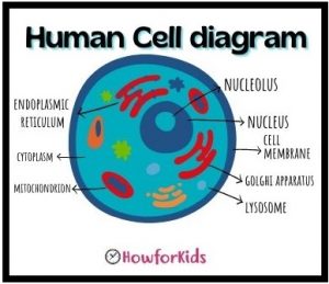
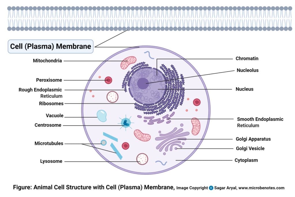
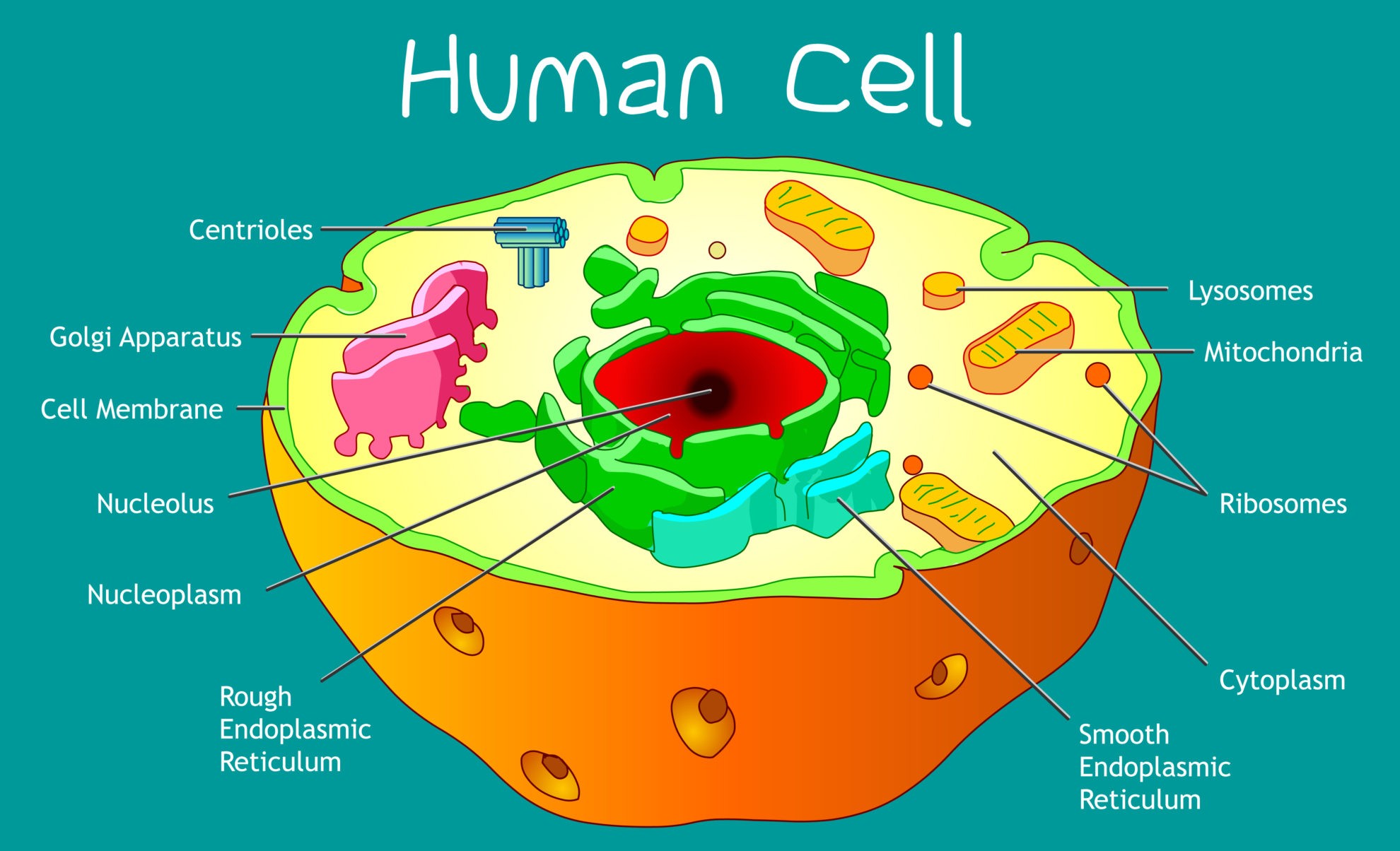
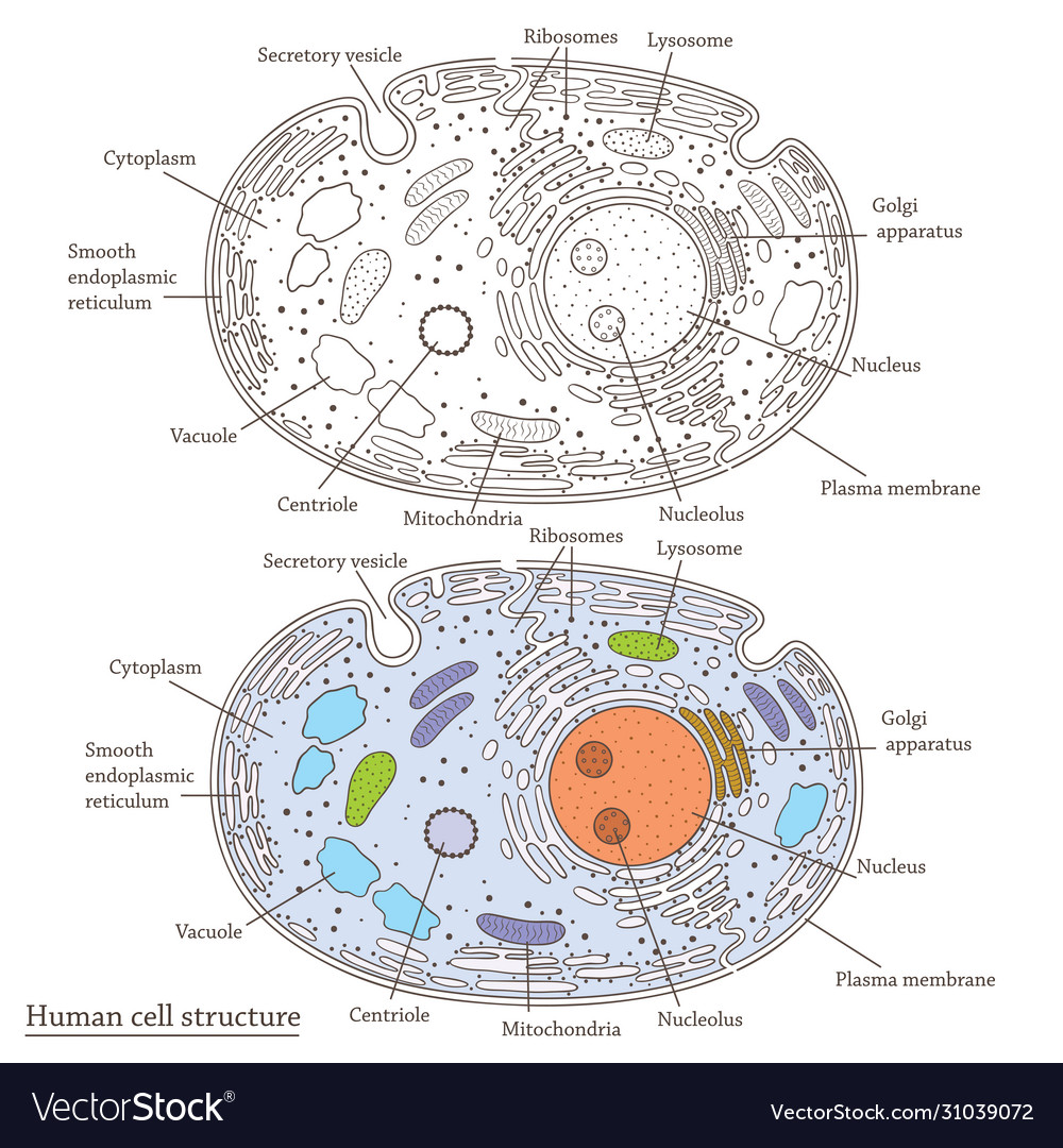
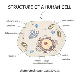
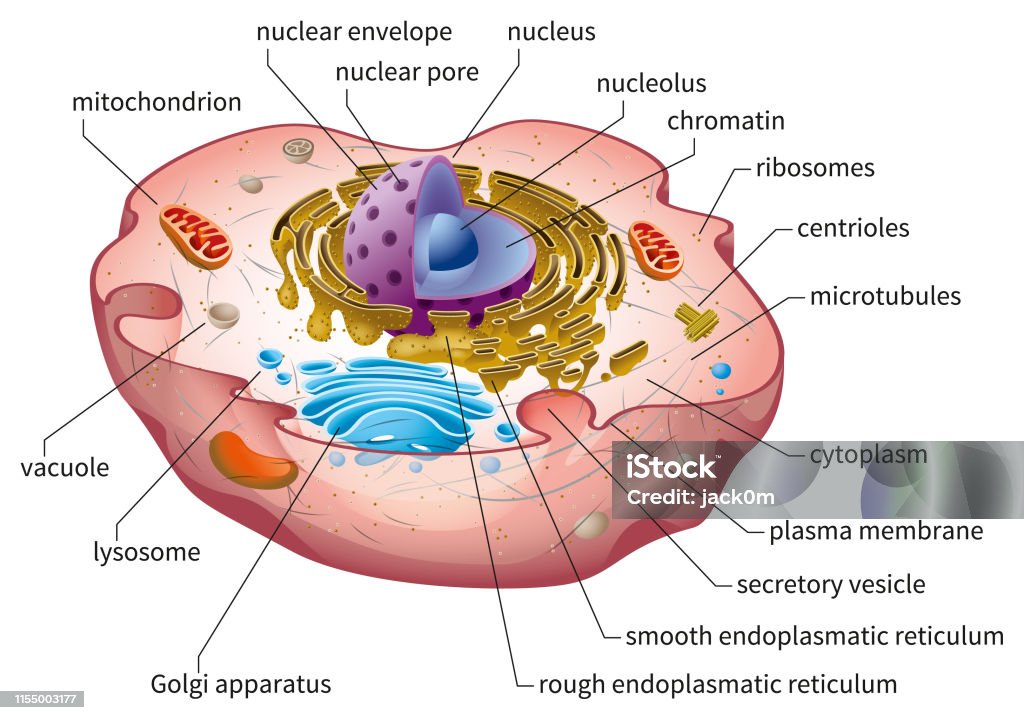
43 diagram of a human cell with labels
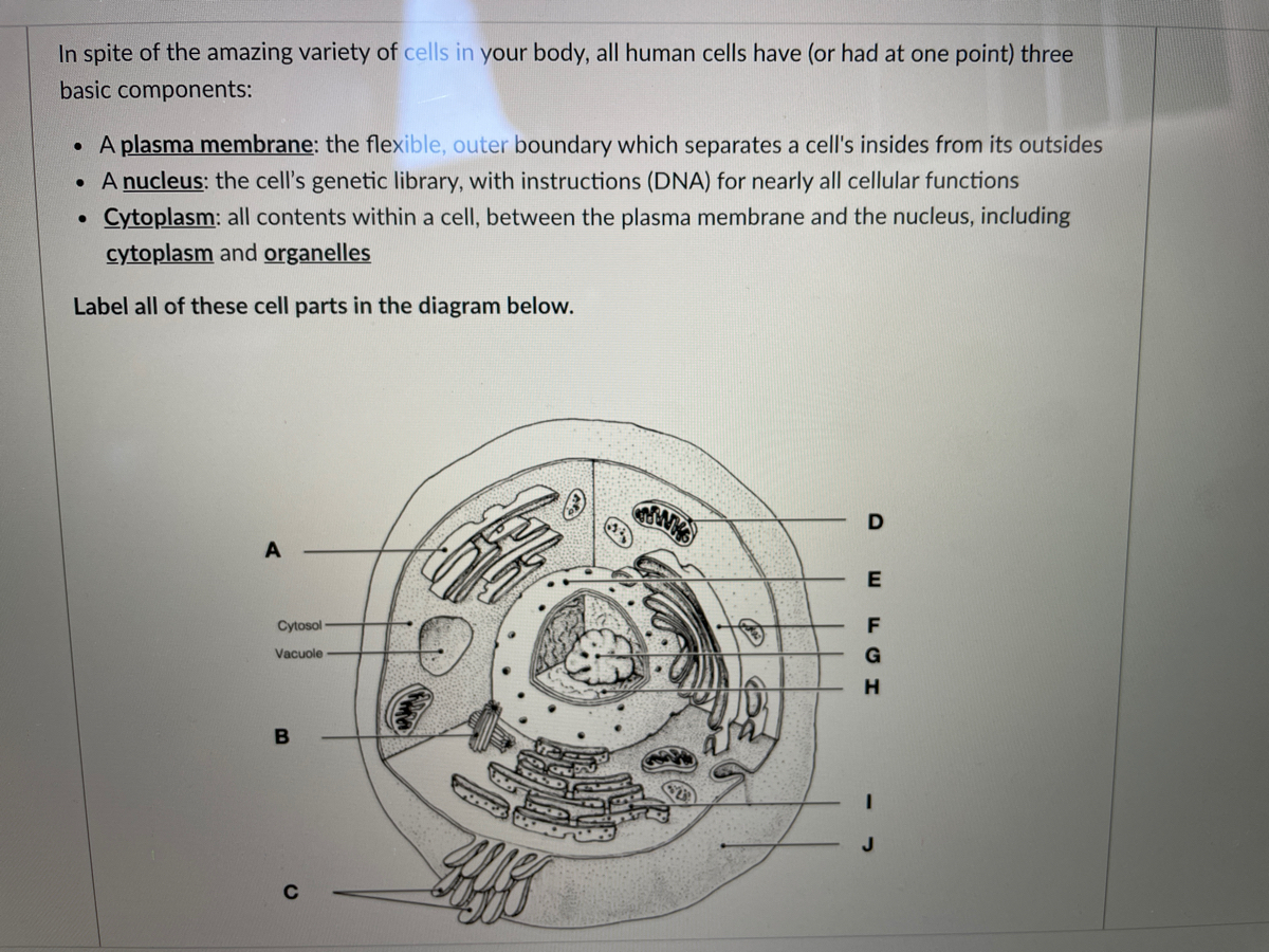
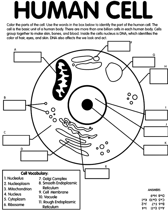
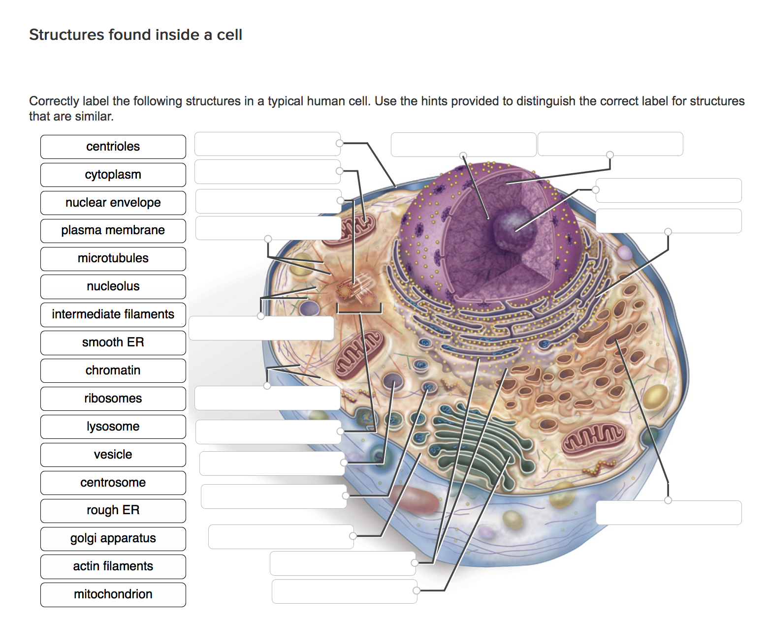
Learn the parts of a cell with diagrams and cell quizzes For this exercise we'll start with an image of a cell diagram ready labeled. Study this and make sure that you're clear about which structure is found where. Cell diagram unlabeled It's time to label the cell yourself! As you fill in the cell structure worksheet, remember the functions of each part of the cell that you learned in the video. Free Anatomy Quiz - The anatomy of the cell - Quiz 1 Study aids. Related quizzes:. Physiology of the cell, Quiz 1 - Now you know the parts of the cell, learn how they function.; The anatomy of bones, Quiz 1 - Learn the anatomy of a human long bone.; The anatomy of muscle, Quiz 1 - How much do you know about the anatomy of a the different muscle types?; Images and pdf's:. Just in case you get tired of looking at the screen we've provided images ...
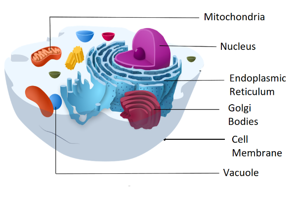
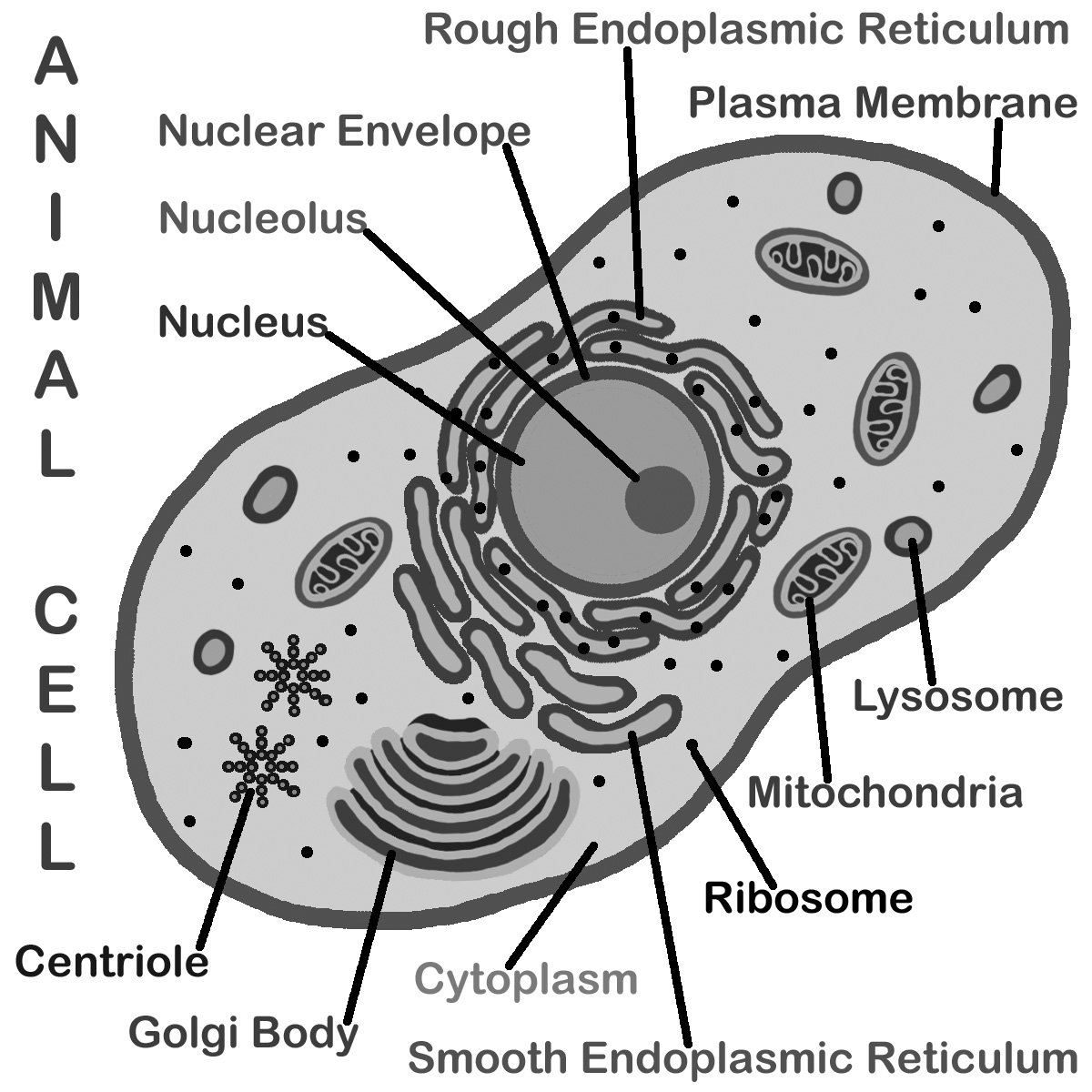
A Labeled Diagram of the Animal Cell and its Organelles A Labeled Diagram of the Animal Cell and its Organelles There are two types of cells - Prokaryotic and Eucaryotic. Eukaryotic cells are larger, more complex, and have evolved more recently than prokaryotes. Where, prokaryotes are just bacteria and archaea, eukaryotes are literally everything else.
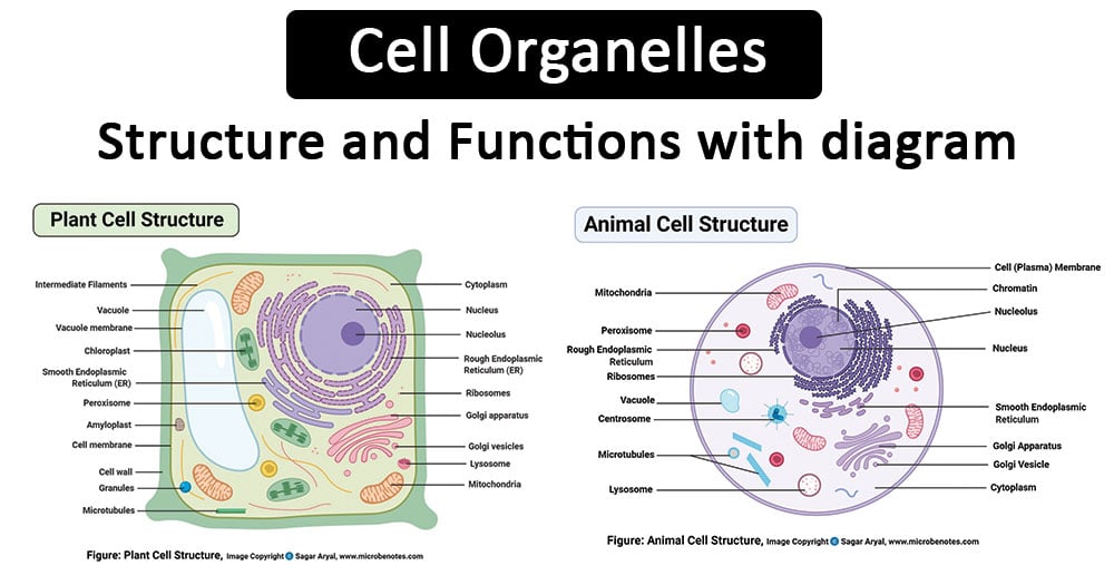
Diagram of a human cell with labels
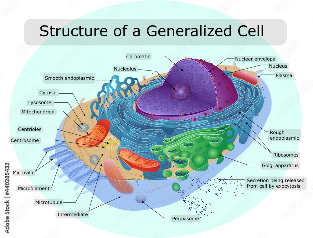
PDF Human Cell Diagram, Parts, Pictures, Structure and Functions Human Cell Diagram, Parts, Pictures, Structure and Functions The cell is the basic functional in a human meaning that it is a self-contained and fully operational living entity. Humans are multicellular organisms with various different types of cells that work together to sustain life. Other non-cellular components in the body include water ... Human Cell Diagram, Parts, Pictures, Structure and Functions Diagram of the human cell illustrating the different parts of the cell. Cell Membrane. The cell membrane is the outer coating of the cell and contains the cytoplasm, substances within it and the organelle. It is a double-layered membrane composed of proteins and lipids. The lipid molecules on the outer and inner part (lipid bilayer) allow it to ... A Labeled Diagram of the Plant Cell and Functions of its Organelles The cell membrane is a thin layer made up of proteins, lipids, and fats. It forms a protective wall around the organelles contained within the cell. It is selectively permeable and thus, regulates the transportation of materials needed for the survival of the organelles of the cell. Function: Protects the cell from its surroundings.
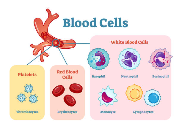
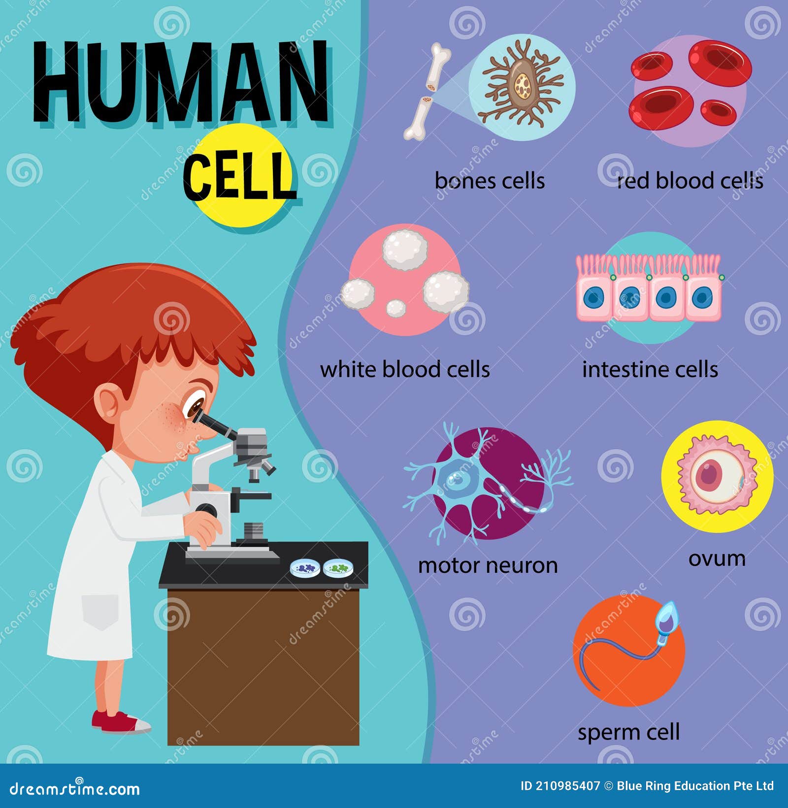
Diagram of a human cell with labels. human cell diagram to label neuroglia cells glial cell types nervous system neurons pns central spinal cns cord satellite schwann function nerve parts myelin axon. 33 Human Cell Diagram To Label - Labels For You duundalleandern.blogspot.com. Unlabelled respiratory system clip art at clker.com. Nucleus cell biology exams. Creating a diagram cell 03 Label the Cell Diagram | Quizlet Start studying 03 Label the Cell. Learn vocabulary, terms, and more with flashcards, games, and other study tools. ... cell diagram. 18 terms. lugo_janet. Sets found in the same folder. 03 Organelle Functions. 14 terms. muskopf1. ... Hole's Human Anatomy and Physiology 13th Edition David N. Shier, Jackie L. Butler, Ricki Lewis. Human Cells Printables and Diagrams - The Successful Homeschool These cells include: leukocytes, haematids, thrombocytes, ovum, sperm, sarcomeres, enterocytes, neurons, osteocytes, hepatocytes. They will learn the parts of a cell thanks to a labeled diagram. They will get to see what blood looks like under a microscope without needing to own a microscope. They get to color a cell and then label the parts. Draw a labelled diagram of human cheek cells. [3 MARKS] Draw a labelled diagram of human cheek cells. [3 MARKS] Solution Squamous epithelium is composed of thin and flat cells, with closely packed nuclei. ∙This type of epithelium is found in the lining of the mouth and nasal cavities, blood vessels, and lymph vessels. Suggest Corrections 43 Similar questions Q.
Human Cell Organelles Labeling Diagram | Quizlet Human Cell Organelles Labeling + − Flashcards Learn Test Match Created by Mackenna_Rios5 Terms in this set (8) Vesicles Transports molecules between organelles and the cell membrane Ribosome Makes Protein Mitochondria Makes ATP Smooth ER Makes lipids and vesicles Lysosomes Breaks down lipids, toxins, bacteria and old cell parts Rough ER How to draw a nerve cell - labeled science diagrams - YouTube Download a free printable outline of this video and draw along with us: you for watching. Please su... 141,840 Labelled Cell Images, Stock Photos & Vectors | Shutterstock 141,840 labelled cell stock photos, vectors, and illustrations are available royalty-free. See labelled cell stock video clips Image type Orientation Color People Artists More Sort by Popular Biology Healthcare and Medical Computing Devices and Phones Technology Animals and Wildlife cell anatomy eukaryote tissue organelle of 1,419 2,894 Nerve Cell Diagram Images, Stock Photos & Vectors - Shutterstock Find Nerve cell diagram stock images in HD and millions of other royalty-free stock photos, illustrations and vectors in the Shutterstock collection. Thousands of new, high-quality pictures added every day.
Cell: Structure and Functions (With Diagram) - Biology Discussion Eukaryotic Cells: 1. Eukaryotes are sophisticated cells with a well defined nucleus and cell organelles. 2. The cells are comparatively larger in size (10-100 μm). 3. Unicellular to multicellular in nature and evolved ~1 billion years ago. 4. The cell membrane is semipermeable and flexible. 5. These cells reproduce both asexually and sexually. Labelled Diagram Of A Human Cell Bone Cell Labeled Diagram Animal Cell ... Oct 7, 2018 - Labelled Diagram Of A Human Cell Bone Cell Labeled Diagram Animal Cell Free Printable To Label. Oct 7, 2018 - Labelled Diagram Of A Human Cell Bone Cell Labeled Diagram Animal Cell Free Printable To Label. Pinterest. Today. Watch. Explore. When autocomplete results are available use up and down arrows to review and enter to select ... A Well-labelled Diagram Of Animal Cell With Explanation - BYJUS Animal cells are eukaryotic cells that contain a membrane-bound nucleus. They are different from plant cells in that they do contain cell walls and chloroplast. The animal cell diagram is widely asked in Class 10 and 12 examinations and is beneficial to understand the structure and functions of an animal. A brief explanation of the different ... Labeled Diagram of the Human Kidney - Bodytomy Labeled Diagram of the Human Kidney The human kidneys house millions of tiny filtration units called nephrons, which enable our body to retain the vital nutrients, and excrete the unwanted or excess molecules as well as metabolic wastes from the body. In addition, they also play an important role in maintaining the water balance of our body.
human cell diagram with labels cell labeled onion epidermal elodea leaf cells labels lab function diagram microscope label salt every manual following slide visible living Lower Leg Bones 1024×1350 | Anatomy System - Human Body Anatomy Diagram anatomysystem.com bones anatomysystem Cell Coloring Page - Coloring Home coloringhome.com
File:Diagram human cell nucleus tr.svg - Wikimedia Commons Description: en: A diagram of a human cell nucleus, with Turkish labels. Translated version of File:Diagram human cell nucleus.svg, originally created and all rights released by Mariana Ruiz (User:LadyofHats).This image is also released to the public domain. az: İnsan hüceyrə nüvəsinin sxematik rəsmi, azərbaycanca yazılı. Source: File:Diagram human cell nucleus.svg
Diagram of human skin structure — Science Learning Hub Diagram of human skin structure. Image. Add to collection. Tweet. Rights: University of Waikato Published 1 February 2011 Size: 100 KB Referencing Hub media. The epidermis is a tough coating formed from overlapping layers of dead skin cells.
Labelled Diagram of Human Eye, Explanation and Function - VEDANTU The human eye is a part of the sensory nervous system. Labeled Diagram of Human Eye The eyes of all mammals consist of a non-image-forming photosensitive ganglion within the retina which receives light, adjusts the dimensions of the pupil, regulates the availability of melatonin hormones, and also entertains the body clock.
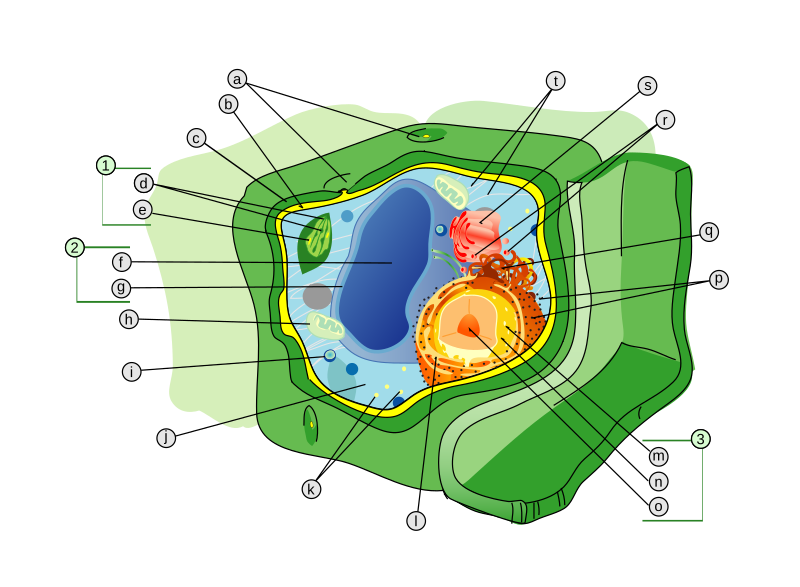
Labeled Plant Cell With Diagrams | Science Trends The parts of a plant cell include the cell wall, the cell membrane, the cytoskeleton or cytoplasm, the nucleus, the Golgi body, the mitochondria, the peroxisome's, the vacuoles, ribosomes, and the endoplasmic reticulum. Parts Of A Plant Cell The Cell Wall Let's start from the outside and work our way inwards.
cell diagram | human cell drawing | anatomy drawing | label cell ... cell diagram | human cell drawing | anatomy drawing | label cell diagram | #shortscell structure:- division :- ...
Diagram of Human Heart and Blood Circulation in It Four Chambers of the Heart and Blood Circulation. The shape of the human heart is like an upside-down pear, weighing between 7-15 ounces, and is little larger than the size of the fist. It is located between the lungs, in the middle of the chest, behind and slightly to the left of the breast bone. The heart, one of the most significant organs ...
human cell diagram to label diagram cell labeled animal human cells anatomy parts chart labelled body labels structure function ANAT2241 Liver, Gallbladder, And Pancreas - Embryology embryology.med.unsw.edu.au histology gallbladder bladder cells gall tissue epithelium human embryology tissues duct normal bile liver pancreas tract atlas slides gastrointestinal cell
Labeled Diagrams of the Human Brain You'll Want to Copy Now - Bodytomy All the functions are carried out without a single glitch and before you even bat an eyelid. The following are the different regions of the human brain and their functions. Labeled Diagrams of the Human Brain Central Core The central core consists of the thalamus, pons, cerebellum, reticular formation and medulla.
Human Cell Diagram Pictures, Images and Stock Photos Diagram of generic plant and animal cells, showing major organelles including nucleus, nucleolus, rough endoplasmic reticulum, smooth endoplasmic reticulum, cell membranes, golgi apparatus, mitochondria, vacuoles, lysosomes, ribosomes, and centrioles. The plant cell obviously also has a cell wall and chloroplasts.
Human cell diagram | Human cell diagram, Cell diagram, Human cell structure With this softly colored felt set you can explore how, where and why this cell works and what each part does. With multiple copies of some parts and a large cell to work will the arrangement possibilities are endless. The labels are felt backed and laminated. These include: Human Cell Golgi apparatus Golgi vesicles Plasma membrane Lysosome….
A Labeled Diagram of the Plant Cell and Functions of its Organelles The cell membrane is a thin layer made up of proteins, lipids, and fats. It forms a protective wall around the organelles contained within the cell. It is selectively permeable and thus, regulates the transportation of materials needed for the survival of the organelles of the cell. Function: Protects the cell from its surroundings.
Human Cell Diagram, Parts, Pictures, Structure and Functions Diagram of the human cell illustrating the different parts of the cell. Cell Membrane. The cell membrane is the outer coating of the cell and contains the cytoplasm, substances within it and the organelle. It is a double-layered membrane composed of proteins and lipids. The lipid molecules on the outer and inner part (lipid bilayer) allow it to ...
PDF Human Cell Diagram, Parts, Pictures, Structure and Functions Human Cell Diagram, Parts, Pictures, Structure and Functions The cell is the basic functional in a human meaning that it is a self-contained and fully operational living entity. Humans are multicellular organisms with various different types of cells that work together to sustain life. Other non-cellular components in the body include water ...







:watermark(/images/watermark_5000_10percent.png,0,0,0):watermark(/images/logo_url.png,-10,-10,0):format(jpeg)/images/overview_image/1032/cDNpPoQ1qy4qxCDShLd5w_histology-eukaryotic-cell_english.jpg)










Post a Comment for "43 diagram of a human cell with labels"